


Research and publish the best content.
Get Started for FREE
Sign up with Facebook Sign up with X
I don't have a Facebook or a X account
Already have an account: Login

Bioscience News - GEG Tech top picks
8.8K views |
+0 today
 Your new post is loading...
 Your new post is loading...
Your new post is loading...
http://www.sciencedirect.com/science/article/pii/S1053811916300891Click here to edit the content
BigField GEG Tech's insight:
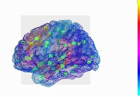
Researchers at CNRS, INSERM, Aix-Marseille University and AP-HM have just created a virtual brain that can reconstitute the brain of a person affected by epilepsy for the first time. From this work we understand better how the disease works and can also better prepare for surgery. These results are published in Neuroimage, on July 28, 2016. 

BigField GEG Tech's insight:
Scientists have created an unprecedented high-resolution map of the brain that reveals structures as small as those found in individual nerve cells. They hope images will enable study of abnormal connections betdepressionween cells present in neurological disorders such as schizophrenia and depression.
|
BigField GEG Tech's insight:
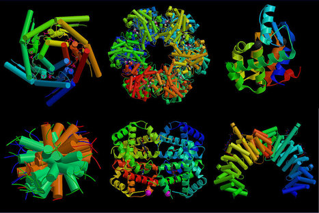
Cells face a daunting task. They have to neatly pack a several meter-long thread of genetic material into a nucleus that measures only five micrometers across. This origami creates spatial interactions between genes and their switches, which can affect human health and disease. Now, an international team of scientists has devised a powerful new technique that 'maps' this three-dimensional geography of the entire genome.
BigField GEG Tech's insight:
A new map of the human brain could be the most accurate yet, as it combines all sorts of different kinds of data. This might finally solve a century of disagreements over the shapes and positions of different brain areas.
|

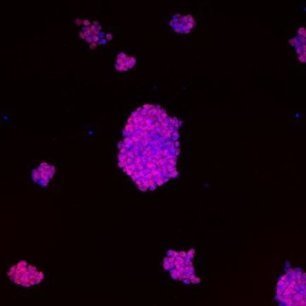


Here, the authors present a comprehensive image-based map of the subcellular protein distribution, the Cell Atlas, built by integrating transcriptomics and antibody-based immunofluorescence microscopy with validation by mass spectrometry.