Host Translocation of Plant Pathogen Effectors
6.9K views | +0 today
Follow
Host Translocation of Plant Pathogen Effectors
Reports of direct microscopic observations of microbial effectors inside pant cells. Some are convincing, others not. You judge and comment!
Your new post is loading...
Scooped by Kamoun Lab @ TSL
Scoop.it!

New Phytologist: Delivery of cytoplasmic and apoplastic effectors from Phytophthora infestans haustoria by distinct secretion pathways (2017)

New Phytologist: Delivery of cytoplasmic and apoplastic effectors from Phytophthora infestans haustoria by distinct secretion pathways (2017) | Host Translocation of Plant Pathogen Effectors | Scoop.it


  • The potato blight pathogen Phytophthora infestans secretes effector proteins that are delivered inside (cytoplasmic) or can act outside (apoplastic) plant cells to neutralize host immunity. Little is known about how and where effectors are secreted during infection, yet such knowledge is essential to understand and combat crop disease.
  • We used transient Agrobacterium tumefaciens-mediated in planta expression, transformation of P. infestans with fluorescent protein fusions and confocal microscopy to investigate delivery of effectors to plant cells during infection.
  • The cytoplasmic effector Pi04314, expressed as a monomeric red fluorescent protein (mRFP) fusion protein with a signal peptide to secrete it from plant cells, did not passively re-enter the cells upon secretion. However, Pi04314-mRFP expressed in P. infestans was translocated from haustoria, which form intimate interactions with plant cells, to accumulate at its sites of action in the host nucleus. The well-characterized apoplastic effector EPIC1, a cysteine protease inhibitor, was also secreted from haustoria. EPIC1 secretion was inhibited by brefeldin A (BFA), demonstrating that it is delivered by conventional Golgi-mediated secretion. By contrast, Pi04314 secretion was insensitive to BFA treatment, indicating that the cytoplasmic effector follows an alternative route for delivery into plant cells.
  • Phytophthora infestans haustoria are thus sites for delivery of both apoplastic and cytoplasmic effectors during infection, following distinct secretion pathways.
No comment yet.
Scooped by Kamoun Lab @ TSL
Scoop.it!

Metabolic priming by a secreted fungal effector. Nature (2011)

Metabolic priming by a secreted fungal effector. Nature (2011) | Host Translocation of Plant Pathogen Effectors | Scoop.it

To localize Cmu1 during biotrophic growth, plants were infected with SG200Dcmu1-cmu1–HA, which carries a cmu1–HA fusion gene inserted in single copy under control of its native promoter. Plants infected with SG200 or with SG200 Pcmu1GFP–HA expressing cytoplasmic green fluorescent protein (GFP) under the cmu1 promoter served as negative controls. Freeze-substituted and resin-embedded sections of maize tissue harvested 3 days after infection with these strains were incubated with anti-HA antibodies and gold markers. Cmu1–HA could be detected inside the fungal hyphae, in the biotrophic interface as well as inside the plant cytoplasm but rarely in the plant cell wall (Fig. 2A and Supplementary Fig. 7). The distribution of gold particles was quantified (Fig. 2B). Gold labelling of plant tissue infected with the parental strain SG200 was negligible (Supplementary Fig. 8), whereas non-secreted GFP–HA was absent from the biotrophic interphase, showed strong accumulation in the fungal cytosol and weak background labelling in the plant cytosol (Supplementary Fig. 9 and Fig. 2B).

No comment yet.
Scooped by Kamoun Lab @ TSL
Scoop.it!

Translocation of Magnaporthe oryzae Effectors into Rice Cells and Their Subsequent Cell-to-Cell Movement. Plant Cell (2010)

Translocation of Magnaporthe oryzae Effectors into Rice Cells and Their Subsequent Cell-to-Cell Movement. Plant Cell (2010) | Host Translocation of Plant Pathogen Effectors | Scoop.it

PWL2 and BAS1 (for biotrophy-associated secreted protein 1), BIC-localized secreted proteins, were translocated into the rice cytoplasm. By contrast, BAS4, which uniformly outlines the IH, was not translocated into the host cytoplasm. Fluorescent PWL2 and BAS1 proteins that reached the rice cytoplasm moved into uninvaded neighbors, presumably preparing host cells before invasion.

Kamoun Lab @ TSL's comment, April 9, 2013 8:04 PM
But this assay is not "pathogen-independent"...
Kamoun Lab @ TSL's comment, April 9, 2013 8:08 PM
Two issues came up in our internal discussions of the maggie RFP-NLS assay:

1/ It would be nice to see a decrease of nuclear signal as you move away from the IH/BIC plant cell. Sometimes there are several cells with stained nuclei - one would expect the signal to become weaker and weaker as the protein gets diluted. Some clear documentation of the weakening of the signal as you move away from the first infected cells would be nice.

2/ A question arose last year about whether the artificial NLS is actually carrying the effector inside the plant cells since there are papers about NLS sequences acting as cell-penetrating peptides.
Mark Farman's comment, April 9, 2013 8:53 PM
There is NO evidence that any Magnaporthe effector translocates across host cell membranes independently of pathogen machinery (nor is there such evidence for oomycetes, for that matter). The Ribot paper is seriously and fundamentally flawed. Actually, I would like to retract my use of the word "translocation" in my earlier comment and replace it with "delivery" because I do not believe that Magnaporthe effectors translocate across membranes at all! Figure that one out...
Scooped by Kamoun Lab @ TSL
Scoop.it!

AY-WB Phytoplasma Secretes a Protein That Targets Plant Cell Nuclei. MPMI (2009)

AY-WB Phytoplasma Secretes a Protein That Targets Plant Cell Nuclei. MPMI (2009) | Host Translocation of Plant Pathogen Effectors | Scoop.it

SAP11, contains an N-terminal SP sequence and a eukaryotic bipartite nuclear localization signal (NLS). SAP11 was detected by immunocytology in nuclei of young sink tissues of China aster plants infected with AY-WB.

No comment yet.
Scooped by Kamoun Lab @ TSL
Scoop.it!

A translocation signal for delivery of oomycete effector proteins into host plant cells. Nature (2007)

A translocation signal for delivery of oomycete effector proteins into host plant cells. Nature (2007) | Host Translocation of Plant Pathogen Effectors | Scoop.it

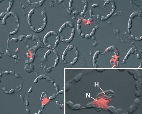
To investigate further the translocation of Avr3a from haustoria into the plant cell, the signal peptide (SP)-RXLR-EER-encoding domain of Avr3a, and a version in which the motifs were replaced with alanine residues (SP-AAAA-AAA) were fused to the amino terminus of the Escherichia coli gusA gene, which encodes β-glucuronidase (GUS), and stably expressed in P. infestans. GusA was chosen because its product is active within plant cells but inactive in the apoplast19, and is thus an ideal reporter for translocation of proteins to the inside of plant cells. SP-RXLR-EER–GUS and SP-AAAA-AAA–GUS transformants were selected that expressed and secreted high levels of active GUS into culture medium (Supplementary Fig. 6). However, when infecting potato, GUS activity was observed only with the SP-RXLR-EER–GUS transformants, and only within plant cells in contact with haustoria (Fig. 3 and Supplementary Fig. 7). No GUS activity was observed within plant cells or in the apoplast in the case of the SP-AAAA-AAA–GUS transformants (Fig. 3 and Supplementary Fig. 7), indicating that GUS was not translocated into the plant cell.

Mark Farman's comment, April 9, 2013 8:08 PM
This is a dead cell. The cytoplasm of a living cell should be distributed around the cell periphery, See the image from the Khang et al. paper.
Kamoun Lab @ TSL's comment, April 9, 2013 8:18 PM
Agreed. Easy to generate such images. See comment in Bozkurt et al. Curr Opin Plant Biol http://kamounlab.dreamhosters.com/pdfs/COPB_2012.pdf
Mark Farman's comment, April 9, 2013 9:29 PM
There is another serious problem with the experiment shown in this figure: In the legend, the authors state that GUS is inactive in the apoplast and they provide a reference. However, the referenced paper deals with GUS that is expressed in plant cells and directed to the apoplast through the secretory pathway. In this case, the GUS is inactivated by glycosylation in the plant ER. Unfortunately, this cannot possibly explain the lack of GUS staining with the apoplastic SP-7A_GUS fusion protein because the SP-RXLR-EER-GUS protein is perfectly visible and had to navigate the same Phytophthora secretory system. In other words, the negative control did not work as it should have done. End result: experiment is invalid.
Scooped by Kamoun Lab @ TSL
Scoop.it!

Ultrastructural changes and immunocytochemical localization of the elicitin quercinin in Quercus robur L. roots infected with Phytophthora quercina. Physiol Mol Plant Pathology (2002)

Ultrastructural changes and immunocytochemical localization of the elicitin quercinin in Quercus robur L. roots infected with Phytophthora quercina. Physiol Mol Plant Pathology (2002) | Host Translocation of Plant Pathogen Effectors | Scoop.it

Immunofluorescence investigations in combination with a specific anti cryptogein antibody clearly showed that the P. quercina elicitin quercinin was located within the hyphal cell wall and seems to be released into invaded cells. In addition, immunogold labelling showed that quercinin was found in the intercellular spaces as well as in penetrated cells.

No comment yet.
Scooped by Kamoun Lab @ TSL
Scoop.it!

Role of the Hrp Pilus in Type III Protein Secretion in Pseudomonas syringae. Science (2001)

Role of the Hrp Pilus in Type III Protein Secretion in Pseudomonas syringae. Science (2001) | Host Translocation of Plant Pathogen Effectors | Scoop.it

We used an in situ immunogold labeling procedure to visualize the extrusion of an effector protein, AvrPto, from the tip of the Hrp pilus, providing direct evidence that a bacterial pilus can function as a conduit for protein delivery.

No comment yet.
Scooped by Kamoun Lab @ TSL
Scoop.it!

A root-knot nematode-secreted protein is injected into giant cells and targeted to the nuclei. New Phytol (2012)

A root-knot nematode-secreted protein is injected into giant cells and targeted to the nuclei. New Phytol (2012) | Host Translocation of Plant Pathogen Effectors | Scoop.it

• Root-knot nematodes (RKNs) are obligate endoparasites that maintain a biotrophic relationship with their hosts over a period of several weeks and induce the differentiation of root cells into specialized feeding cells. Nematode effectors synthesized in the oesophageal glands and injected into the plant tissue through the syringe-like stylet certainly play a central role in these processes.
• In a search for nematode effectors, we used comparative genomics on expressed sequence tag (EST) datasets to identify Meloidogyne incognita genes encoding proteins potentially secreted upon the early steps of infection.
• We identified three genes specifically expressed in the oesophageal glands of parasitic juveniles that encode predicted secreted proteins. One of these genes, Mi-EFF1 is a pioneer gene that has no similarity in databases and a predicted nuclear localization signal. We demonstrate that RKNs secrete Mi-EFF1 within the feeding site and show Mi-EFF1 targeting to the nuclei of the feeding cells.
• RKNs were previously shown to secrete proteins in the apoplasm of infected tissues. Our results show that nematodes sedentarily established at the feeding site also deliver proteins within plant cells through their stylet. The protein Mi-EFF1 injected within the feeding cells is targeted at the nuclei where it may manipulate nuclear functions of the host cell.

No comment yet.
Scooped by Kamoun Lab @ TSL
Scoop.it!

A Secreted Fungal Effector of Glomus intraradices Promotes Symbiotic Biotrophy. Current Biology (2011)

A Secreted Fungal Effector of Glomus intraradices Promotes Symbiotic Biotrophy. Current Biology (2011) | Host Translocation of Plant Pathogen Effectors | Scoop.it

We show that Glomus intraradices secretes a protein, SP7, that interacts with the pathogenesis- related transcription factor ERF19 in the plant nucleus. SP7-mRFP is secreted by a Magnaporthe oryzae GFP-expressing strain, translocated into the onion cell, and localized to the nucleus. No red nuclei were detected when the infection assay was performed using the GFP control strain (Guy11-GFP). Images show maximal projections of several confocal planes. Scale bars represent 10 mm.

Gregor Langen's comment, April 11, 2013 3:57 AM
I don't expect big differences in onion skin response to pathogenic or mutualistic symbionts regarding nuclear movemnt. Eveln mechanical stimulation causes movemenr of the nucleus: Local mechanical stimulation induces components of the pathogen defense response in parsley. Gus-Mayer et al. PNAS 1998 95(14):8398-403.
But I agree that DAPI staining would have helped to clarify identitiy of the structures visible. And yes, halos can be seen
Gregor Langen's comment, April 11, 2013 4:02 AM
and I agree that a red halo itself doesn't proof that its identical to the nucleus.
Mark Farman's comment, April 27, 2013 10:50 PM
Even if the onion nuclei are beneath the appressoria, the authors have not established that the fluorescence is in the nucleus, as opposed to being on the surface of the cuticle.
Scooped by Kamoun Lab @ TSL
Scoop.it!

A Secreted Effector Protein of Laccaria bicolor Is Required for Symbiosis Development. Current Biology (2011)

A Secreted Effector Protein of Laccaria bicolor Is Required for Symbiosis Development. Current Biology (2011) | Host Translocation of Plant Pathogen Effectors | Scoop.it

MiSSP7 was computationally predicted to be secreted into the plant apoplastic space [3]. Its localization after secretion was further investigated here. Using immunofluorometric labeling, we found that MiSSP7 enters plant cells and accumulates in the plant nuclei (Figure 1C).

No comment yet.
Scooped by Kamoun Lab @ TSL
Scoop.it!

Internalization of Flax Rust Avirulence Proteins into Flax and Tobacco Cells Can Occur in the Absence of the Pathogen. Plant Cell (2010)

Internalization of Flax Rust Avirulence Proteins into Flax and Tobacco Cells Can Occur in the Absence of the Pathogen. Plant Cell (2010) | Host Translocation of Plant Pathogen Effectors | Scoop.it

We show by immunolocalization that the flax rust AvrM protein is secreted from haustoria during infection and accumulates in the haustorial wall. Five days after inoculation, the AvrM protein was also detected within the cytoplasm of a proportion of plant cells containing haustoria, confirming its delivery into host cells during infection.

Mark Farman's comment, April 9, 2013 8:25 PM
Despite the authors assertion that there is no labeling of uninfected cells - there is. Therefore, the fluorescence in the cytoplasmic rings is probably background. Problem is there were no negative (non infected, pre-immune serum) controls in this paper, so we can't tell either way. The fluorescence patterns in the middle panel on the righthand side do not coincide with the cytoplasmic boundaries, indicating a problem with fixation. Most likely protein leached out from the haustoria into the spaces that should be vacuolar.
Scooped by Kamoun Lab @ TSL
Scoop.it!

Identification of a Protein from Rust Fungi Transferred from Haustoria into Infected Plant Cells. MPMI (2005)

Identification of a Protein from Rust Fungi Transferred from Haustoria into Infected Plant Cells. MPMI (2005) | Host Translocation of Plant Pathogen Effectors | Scoop.it
Rust transferred protein 1 from Uromyces fabae (Uf-RTP1p) was not only detected in the host parasite interface, the extrahaustorial matrix, but also inside infected plant cells by immunofluorescence and electron microscopy. Uf-RTP1p does not exhibit any similarity to sequences currently listed in the public databases. However, we identified a homolog of Uf-RTP1p in the related rust fungus Uromyces striatus (Us-RTP1p). The localization of Uf-RTP1p and Us-RTP1p inside infected plant cells was confirmed, using four independently raised polyclonal antibodies. Depending on the developmental stage of haustoria, Uf-RTP1p was found in increasing amounts in host cells, including the host nucleus.
Mark Farman's comment, April 9, 2013 8:13 PM
This staining is NOT in the cytoplasm - it is in the extrahaustorial matrix. Also where's the evidence that the stained structure on the bottom right is a nucleus?
Eric Kemen's comment, May 3, 2013 11:27 AM
I agree, it’s not easy form this picture to see that there is a nucleus underneath the fluorescence signal if one hasn’t seen the DIC picture. But have a look at figure 3 of the same paper where an independent antibody was used in combination with a nuclear stain, to show colocalization of RTP1p signal and DNA staining. In addition, immuno electron microscopy after high pressure freezing shows a signal for RTP1p localization in the cytoplasm (figure 2).
Kamoun Lab @ TSL's comment, May 17, 2013 10:11 AM
Not much mention of nuclear localization in this follow-up paper http://sco.lt/5jAe01
Suggested by dromius
Scoop.it!

Type III-dependent translocation of the Xanthomonas AvrBs3 protein into the plant cell. Mol Microbiology (2002)

Type III-dependent translocation of the Xanthomonas AvrBs3 protein into the plant cell. Mol Microbiology (2002) | Host Translocation of Plant Pathogen Effectors | Scoop.it

Using an immunocytochemical approach, we demonstrate that the type III effector AvrBs3 from Xanthomonas campestris pv. vesicatoria localizes to nuclei of infected pepper leaves. Importantly, AvrBs3 translocation was observed in situ in native tissues of susceptible and resistant plants. AvrBs3 was detected in the nucleus as soon as 4 h post infection, which was dependent on a functional TTSS and the putative translocator HrpF. N-terminal AvrBs3 deletion derivatives are no longer secreted by the TTSS in vitro and could not be detected inside the host cells, suggesting that the N-terminus of AvrBs3 is important for secretion.

No comment yet.