


 Your new post is loading...

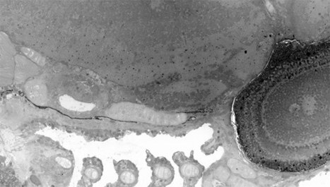

Electron microscopes can produce incredibly detailed and even 3D views of sub-cellular structures, but often at the cost of losing the bigger picture. Researchers at Leiden University in the Netherlands, however, have leveraged a technique called virtual nanoscopy that enables researchers to observe the whole of a cell and its intricate details in a single image.
With the method, the team stitches together nanometer resolution photographs of what's gone under the scope to create a map with adjustable zoom a la Google Maps. Their study created a 281-gigapixel image (packed with 16 million pixels per inch) of a 1.5-millimeter-long zebrafish embryo.
Via Anne Osterrieder, Dr. Stefan Gruenwald


