Your new post is loading...

|
Scooped by
Juan Lama
|
An outbreak of H5N1 highly pathogenic influenza A virus (HPIAV) has been detected in dairy cows in the United States. Influenza A virus (IAV) is a negative-sense, single-stranded, RNA virus that has not previously been associated with widespread infection in cattle. As such, cattle are an extremely under-studied domestic IAV host species. IAV receptors on host cells are sialic acids (SAs) that are bound to galactose in either an α2,3 or α2,6 linkage. Human IAVs preferentially bind SA-α2,6 (human receptor), whereas avian IAVs have a preference for α2,3 (avian receptor). The avian receptor can further be divided into two receptors: IAVs isolated from chickens generally bind more tightly to SA-α2,3-Gal-β1,4 (chicken receptor), whereas IAVs isolated from duck to SA-α2,3-Gal-β1,3 (duck receptor). We found all receptors were expressed, to a different degree, in the mammary gland, respiratory tract, and cerebrum of beef and/or dairy cattle. The duck and human IAV receptors were widely expressed in the bovine mammary gland, whereas the chicken receptor dominated the respiratory tract. In general, only a low expression of IAV receptors was observed in the neurons of the cerebrum. These results provide a mechanistic rationale for the high levels of H5N1 virus reported in infected bovine milk and show cattle have the potential to act as a mixing vessel for novel IAV generation. Preprint in bioRxiv (May 3, 2024): https://doi.org/10.1101/2024.05.03.592326
|
|
Scooped by
Juan Lama
|
University of Sydney scientists have discovered a protein in the lung that blocks SARS-CoV-2 infection and forms a natural protective barrier in the human body. This protein, the leucine-rich repeat-containing protein 15 (LRRC15), is an inbuilt receptor that binds the SARS-CoV-2 virus without passing on the infection. The research opens up an entirely new area of immunology research around LRRC15 and offers a promising pathway to develop new drugs to prevent viral infection from coronaviruses like COVID-19 or deal with fibrosis in the lungs. The study has been published in the journal PLOS Biology. It was led by Professor Greg Neely with his team members Dr Lipin Loo, a postdoctoral researcher, and PhD student Matthew Waller at the Charles Perkins Centre and the School of Life and Environmental Sciences. The University study is one of three independent papers that reveal this specific protein’s interaction with COVID-19. “Alongside two other groups, one at Oxford, the other at Brown and Yale in the USA, we found a new receptor in the LRRC15 protein that can stop SARS-CoV-2. We found that this new receptor acts by binding to the virus and sequestering it which reduces infection,” Professor Neely said. “For me, as an immunologist, the fact that there's this natural immune receptor that we didn't know about, that's lining our lungs and blocks and controls virus, that's crazy interesting. “We can now use this new receptor to design broad acting drugs that can block viral infection or even suppress lung fibrosis.” What is LRRC15? The COVID-19 virus infects humans by using a spike protein to attach to a specific receptor in our cells. It primarily uses a protein called the angiotensin-converting enzyme 2 (ACE2) receptor to enter human cells. Lung cells have high levels of ACE2 receptors, which is why the COVID-19 virus often causes severe problems in this organ of infected people. Like ACE2, LRRC15 is a receptor for coronavirus, meaning the virus can bind to it. But unlike ACE2, LRRC15 does not support infection. It can, however, stick to the virus and immobilise it. In the process, it prevents other vulnerable cells from becoming infected. “We think it acts a bit like Velcro, molecular Velcro, in that it sticks to the spike of the virus and then pulls it away from the target cell types,” Dr Loosaid. “Basically, the virus is coated in the other part of the Velcro, and while it's trying to get to the main receptor, it can get caught up in this mesh of LRRC15,” Mr Waller said. LRRC15 is present in many locations such as lungs, skin, tongue, fibroblasts, placenta and lymph nodes. But the researchers found human lungs light up with LRRC15 after infection. “When we stain the lungs of healthy tissue, we don't see much of LRRC15, but then in COVID-19 lungs, we see much more of the protein,” Dr Loosaid. “We think this newly identified protein could be part of our body’s natural response to combating the infection creating a barrier that physically separates the virus from our lung cells most sensitive to COVID-19.” Implications of the research “When we studied how this new receptor works, we found that this receptor also controls antiviral responses, as well as fibrosis, and could link COVID-19 infection with lung fibrosis that occurs during long COVID,” Mr Waller said. “Since this receptor can block COVID-19 infection, and at the same time activate our body’s anti-virus response, and suppress our body’s fibrosis response, this is a really important new gene,” Professor Neely said. “This finding can help us develop new antiviral and antifibrotic medicines to help treat pathogenic coronaviruses, and possibly other viruses or other situations where lung fibrosis occurs. “For fibrosis, there are no good drugs: for example, idiopathic pulmonary fibrosis is currently untreatable.” Fibrosis is a condition in which lung tissue becomes scarred and thickened, causing breathing difficulties. COVID-19 can cause inflammation and damage to the lungs, leading to fibrosis. The authors said they are developing two strategies against COVID-19 using LRRC15 that could work across multiple variants – one which targets the nose as a preventative treatment, and another aimed at the lungs for serious cases. The researchers also said that the presence or lack of LRRC15, which is involved in lung repair, is an important indication of how severe a COVID-19 infection might become. “A group at Imperial College London independently found that absence of LRRC15 in the blood is associated with more severe COVID, which supports what we think is happening.” Dr Loo said. “If you have less of this protein, you likely have serious COVID. If you have more of it, your COVID is less severe. “We are now trying to understand exactly why this is the case.” The research involved screening human cell cultures for genes and investigating the lungs of human COVID-19 patients. Findings published in Plos Biology (Feb. 9, 2023): https://doi.org/10.1371/journal.pbio.3001967
|
|
|
Scooped by
Juan Lama
|
In Europe, the pandemic triggered in 2020 by the SARS-CoV-2 coronavirus is now largely under control. But why this virus is able to spread so efficiently remains unclear. A team of researchers led by Dr. Simone Backes, Dr. Gerti Beliu and Prof. Dr. Markus Sauer of the Julius Maximilians University of Würzburg (JMU) has now shown in a publication in "Angewandte Chemie" that some previous assumptions need to be reconsidered. For example, the virus does not bind with several surface proteins simultaneously to several receptors of the cell to be infected. This assumption has previously been an attempt to explain how viruses increase their infectivity. Binding to a single receptor also does not lead to the subsequent docking of further receptors to the virus. The Würzburg research group has now provided evidence that a single virus binds to a single receptor, opening the door for a highly efficient infection. What could only be speculated about SARS-CoV-2 carries an average of 20 - 40 spike proteins on its surface. With these, it binds to ACE2 receptors in the membrane of its target cells, for example in the nose and throat of humans. When these receptors are blocked with antibodies, the cell can no longer be infected. Making the ACE2 receptors and their interaction with the viral spike proteins visible microscopically has not been possible so far. Therefore, much was left to speculation - such as whether the viruses bind to multiple receptors with multiple spikes to facilitate entry into the cell. It was also considered that the receptors are present in the membrane in pairs or groups of three rather, so that they can bind more efficiently to the trimeric spike proteins. Or that they are only combined into such groups after binding to a spike protein. Both depend strongly on the density of the ACE2 receptors in the membrane. Super-resolution microscopy made it clear The Würzburg researchers wanted to elucidate this mystery: They labeled antibodies with dyes to make the receptors visible and countable. To do this, they used various cell lines that are used as model systems for SARS-CoV infection, and the single-molecule sensitive super-resolution microscopy method dSTORM, developed in Markus Sauer's research group. It turned out that Vero cells, for example, which are often used as a model for SARS-CoV-2 infection, only have one to two ACE2 receptors per square micrometre of cell membrane. This is very few: "In other membrane receptors, this number is often between 30 and 80," Sauer added. "The average distance between neighbouring ACE2 receptors is about 500 nanometres. It is thus much larger than a virus particle, which measures only 100 nanometres," says Backes. The idea that a virus particle with multiple spike proteins can bind to multiple receptors simultaneously is therefore very unlikely, she adds. ACE2 receptors are always single The following open question: Are the receptors also present as pairs or groups of three in the membrane? "No. They only occur there singly. And it stays that way even when a viral spike protein has bound to them," says Beliu, group leader at the Rudolf Virchow Center. For an infection, it is sufficient if a single spike binds to a single receptor. With these results, the JMU team was able to disprove many of the original hypotheses about the interaction of viral particles with multiple ACE2 receptors. It also showed that host cells with higher ACE2 expression are more easily to infect, as expected. However, the lipid composition of the membrane and other factors also influence infection efficiency. What is next? The JMU team wants to gather as much detailed knowledge as possible about the cell entry mechanism of coronaviruses in order to better understand the infection process. This could ultimately contribute to better prevention and the development of better drugs against COVID-19. Next, the Würzburg researchers want to analyse the entry mechanism with high-resolution light sheet microscopy. Published March 2023: https://doi.org/10.1002/anie.202300821
|
|
Scooped by
Juan Lama
|
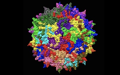
Researchers have identified a novel cellular entry factor for adeno-associated virus vector (AAV) types -- the most commonly used viral vectors for in vivo gene therapy. The researchers identified that GPR108, a G protein-coupled receptor, served as a molecular 'lock' to the cell. The discovery could one day enable scientists to better direct AAV gene transfers to specific tissues. The researchers identified that GPR108, a G protein-coupled receptor, served as a molecular 'lock' to the cell. GPR108 is required for most AAVs, including those used in approved gene therapies, to gain access to the cell. As gaining cellular access is a critical step in delivering gene therapy, this discovery may provide a crucial piece of information that could one day enable scientists to better explain, predict, and ultimately, direct AAV gene transfers to specific tissues. The study was recently published in Molecular Therapy. "For years we have known that AAV gene transfer is highly effective, but we have yet to learn how that is achieved and why some AAV types function differently than others," said senior study author Luk Vandenberghe, PhD, Director of the Grousbeck Gene Therapy Center at Mass. Eye and Ear and Associate Professor of Ophthalmology at Harvard Medical School. "We identified a molecular 'lock' to the cell that allows AAV vectors carrying the appropriate 'key' to gain access to the cell. This finding may enable scientists to better direct AAV gene transfers to targeted cell tissues, in order to treat specific genetic diseases." Multiple AAV types are in clinical trials for diseases affecting the eye, muscles, and neurons. Luxturna™ and Zolgensma™, both recently approved by the U.S. Food and Drug Administration, are AAV gene therapy products for a form of blindness and neuromuscular disease. Yet, the exact mechanism by which this novel class of medicine accomplishes gene transfer has remained poorly understood. Published in Molecular Therapy (November 13, 2019): https://doi.org/10.1016/j.ymthe.2019.11.005
|