Plant Cell Biology and Microscopy
6.3K views | +0 today
Follow
Plant Cell Biology and Microscopy
Methods and Tools for Plant Cell Biology
Curated by dromius
Your new post is loading...
Scooped by dromius
Scoop.it!

J. Exp. Bot. - Live imaging of intra- and extracellular pH in plants using pHusion, a novel genetically encoded biosensor

J. Exp. Bot. - Live imaging of intra- and extracellular pH in plants using pHusion, a novel genetically encoded biosensor | Plant Cell Biology and Microscopy | Scoop.it

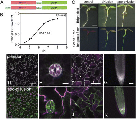
Krag Gjetting et al, 2012


pHusion is especially designed for apoplastic pH measurements. It was constitutively expressed in Arabidopsis and targeted for expression in either the cytosol or the apoplast including intracellular compartments. pHusion consists of the tandem concatenation of enhanced green fluorescent protein (EGFP) and monomeric red fluorescent protein (mRFP1), and works as a ratiometric pH sensor.


No comment yet.
Scooped by dromius
Scoop.it!

BioTechniques - Visualization of cofilin-actin and Ras-Raf interactions by bimolecular fluorescence complementation assays using a new pair of split Venus fragments

BioTechniques - Visualization of cofilin-actin and Ras-Raf interactions by bimolecular fluorescence complementation assays using a new pair of split Venus fragments | Plant Cell Biology and Microscopy | Scoop.it

This article tested different breakpoints of the fluorescent protein Venus. 44 positions resulted in self-reassembly. Others only asssembled when interacting proteins were fused.


A new pair of split Venus fragments, Venus (1–210) fused upstream of cofilin and Venus (210–238) fused downstream of actin, was the most effective combination for visualizing the specific interaction between cofilin and actin in living cells.

No comment yet.
Scooped by dromius
Scoop.it!

Neuron - GFP Reconstitution Across Synaptic Partners (GRASP) Defines Cell Contacts and Synapses in Living Nervous Systems

Neuron - GFP Reconstitution Across Synaptic Partners (GRASP) Defines Cell Contacts and Synapses in Living Nervous Systems | Plant Cell Biology and Microscopy | Scoop.it

We have developed GRASP, a system to label membrane contacts and synapses between two cells ... Two complementary fragments of GFP are expressed on different cells, tethered to extracellular domains of transmembrane carrier proteins. When the complementary GFP fragments are fused to ubiquitous transmembrane proteins, GFP fluorescence appears uniformly along membrane contacts between the two cells. When one or both GFP fragments are fused to synaptic transmembrane proteins, GFP fluorescence is tightly localized to synapses. GRASP marks known synaptic contacts ... and can uncover new information about synaptic locations as confirmed by electron microscopy. GRASP may prove particularly useful for defining connectivity in complex systems.

No comment yet.
Scooped by dromius
Scoop.it!

mEosFP-Based Green-to-Red Photoconvertible Subcellular Probes for Plants

mEosFP-Based Green-to-Red Photoconvertible Subcellular Probes for Plants | Plant Cell Biology and Microscopy | Scoop.it

Photoconvertible fluorescent proteins (FPs) are recent additions to the biologists’ toolbox for understanding the living cell. Like green fluorescent protein (GFP), monomeric EosFP is bright green in color but is efficiently photoconverted into a red fluorescent form using a mild violet-blue excitation. Here, we report mEosFP-based probes that localize to the cytosol, plasma membrane invaginations, endosomes, prevacuolar vesicles, vacuoles, the endoplasmic reticulum, Golgi bodies, mitochondria, peroxisomes, and the two major cytoskeletal elements, filamentous actin and cortical microtubules.

No comment yet.
Scooped by dromius
Scoop.it!

Plant Cell - New Technologies for 21st Century Plant Science

In 2009, a committee of the National Academy highlighted the “understanding of plant growth” as one of the big challenges for society and part of a new era which they termed “new biology.” The aim of this article is to identify how new technologies can and will transform plant science to address the challenges of new biology. We assess where we stand today regarding current technologies, with an emphasis on molecular and imaging technologies, and we try to address questions about where we may go in the future and whether we can get an idea of what is at and beyond the horizon.

No comment yet.
Scooped by dromius
Scoop.it!

The LANL GFP Toolbox

The GFP Toolbox includes four GFP-based tools: Folding Reporter, SuperFolder,
Insertion, and Split GFP. This suite enables researchers to perform experiments
impossible to do with conventional GFPs. The toolbox is simple to use, versatile,
and each component has only one moving part. The toolbox includes genetically
encoded tags as short as 15 amino acids, with little or no effect on passenger
protein localization, behavior, folding, or solubility.

No comment yet.
Scooped by dromius
Scoop.it!

Protein mislocalization in plant cells using a GFP-binding chromobody

Protein mislocalization in plant cells using a GFP-binding chromobody | Plant Cell Biology and Microscopy | Scoop.it

Schornack et al, 2009, Plant Journal


A key challenge in cell biology is to directly link protein localization to function. The green fluorescent protein (GFP)-binding protein, GBP, is a 13-kDa soluble protein derived from a llama heavy chain antibody that binds with high affinity to GFP as well as to some GFP variants such as yellow fluorescent protein (YFP). A GBP fusion to the red fluorescent protein (RFP), a molecule termed a chromobody, was previously used to trace in vivo the localization of various animal antigens. In this study, we extend the use of chromobody technology to plant cells and develop several applications for the in vivo study of GFP-tagged plant proteins.

No comment yet.
Scooped by dromius
Scoop.it!

Immunolabeling artifacts and the need for live-cell imaging : Nature Methods

Immunolabeling artifacts and the need for live-cell imaging : Nature Methods | Plant Cell Biology and Microscopy | Scoop.it

In this Perspective the authors highlight and discuss the artifacts that can arise when using immunolabeling to examine protein localization

No comment yet.