NeuroImmunology
8.2K views | +0 today
Follow
 
Scooped by Gilbert C FAURE from Virus World
onto NeuroImmunology
Scoop.it!

HIV Hides Out In Brain-Resident Immune Cells

HIV Hides Out In Brain-Resident Immune Cells | NeuroImmunology | Scoop.it

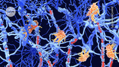
Studies in living brain tissue found that specialized immune cells in the brain can harbor latent but replication-competent HIV.  As a part of its life cycle, human immunodeficiency virus-1 (HIV-1) inserts a copy of its DNA into human immune cells. Some of these newly infected immune cells can then transition into a dormant, latent state for a long period of time, which is known as HIV latency. Although current antiretroviral therapy (ART) against HIV can successfully block the virus from replicating further, it cannot eradicate latent HIV. If treatment is discontinued, the virus can rebound from latency and reignite the progression of HIV infection to AIDS. Scientists at the HIV Cure Center at the UNC School of Medicine, University of California, San Diego (UCSD), Emory University, and the University of Pennsylvania, have been searching for where exactly these latent cells are hiding in the body. Their newly reported studies indicate that brain microglial (BM)—specialized brain-resident immune cells with a decade-long lifespan—can serve as a stable viral reservoir for latent HIV. “We now know that microglial cells serve as a persistent brain reservoir,” said Yuyang Tang, PhD, assistant professor of medicine in the division of infectious diseases and member of the UNC HIV Cure Center. “This had been suspected in the past, but proof in humans was lacking. Our method for isolating viable brain cells provides a new framework for future studies on reservoirs of the central nervous system, and, ultimately, efforts towards the eradication of HIV.” Tang is first author of the team’s published paper in The Journal of Clinical Investigation, which is titled, “Brain microglia serve as persistent HIV reservoir despite durable antiretroviral therapy.”


HIV is a tricky virus to study. During infection, the virus specifically targets CD4+ lymphocytes which are the key coordinators of the immune response. Over time, the virus kills enough CD4+ cells to cause immunodeficiency. Past research has shown that latent HIV can hide within a few of the surviving CD4+ T cells throughout the body and the bloodstream. However, it’s been suspected that there are other viral reservoirs hidden within the central nervous system (CNS) in people with HIV who are receiving effective ART. But as the authors noted, “… rigorous evidence of viral persistence in the CNS cells of humans on durable suppressive ART is incomplete … Brain microglia (MG) may serve as a human immunodeficiency virus 1 (HIV) reservoir and ignite rebound viremia following cessation of antiretroviral therapy (ART), but they have yet to be proven to harbor replication-competent HIV. Unlike peripheral blood cells, it is extremely difficult to access and analyze brain tissues for the study of HIV reservoirs. Since these types of cells cannot be safely sampled in people taking ART, the potential viral reservoir in the brain has remained an enigma for many years. For their reported research the team first studied the brains of macaques infected with simian immunodeficiency virus (SIV), a virus that is closely related to HIV, from the Yerkes National Primate Research Center at Emory University to get a better understanding of how to extract and purify viable cells from primate brain tissue. The researchers used physical separation techniques and antibodies to selectively remove cells that were expressing microglial surface markers. Then, they isolated and separated the highly pure brain myeloid cells (BrMCs) from the CD4+ cells that were passing through the brain tissue. Using these techniques, researchers then obtained samples that were donated by HIV+ people (people with HIV; PWH) who were enrolled in “The Last Gift” Study at UCSD. As a part of this unique and important effort, altruistic HIV+ people, who are taking ART but suffering from other terminal illnesses, will their bodies to further the HIV research project.


“ … we first developed protocols to isolate highly pure populations of BrMCs and MG from the tissues of nonhuman primates (NHPs),” the authors explained. “We then adapted these protocols to the study of human brain tissues containing large numbers of viable cells after rapid autopsy to explore whether human BrMCs produce replication-competent HIV.” Co-author David Margolis, MD, the Sarah Kenan distinguished professor of medicine, microbiology & immunology, and epidemiology, further noted, “The samples are from people living with HIV, who are on therapy but facing a fatal disease of some kind. They were willing to not just donate their bodies to science, but also participate in the research program in the months leading up to their death. It’s an extraordinary program that made this critical research possible.” The scientists’ findings confirmed that MGs from an individual with HIV, being treated using ART, harbored replication-competent HIV. They acknowledged that although their study was limited by the small number of available samples from human donors on ART, they believe that the findings are consistent with NHP studies. “Our observations support the concept that brain MG are a stable reservoir of quiescent infection and may be a source of viral rebound upon treatment interruption,” they concluded. “Future efforts to clear HIV infection will have to include assessments of the persistence of HIV within CNS MG.” Now that the researchers know that latent HIV can take refuge in microglial cells in the brain, they are now considering plans to target this type of reservoir. Since latent HIV in the brain is radically different from the virus in the periphery, researchers believe that it has adapted special characteristics to replicate in the brain.


Reporting in their paper, the team noted, “Phenotyping studies characterized brain-derived virus as macrophage tropic based on the ability of the virus to infect cells expressing low levels of CD4. The lack of genetic diversity in virus from the brain suggests that this macrophage-tropic lineage quickly colonized brain regions.” NF-κB signaling is one of the critical signaling pathways that control HIV expression elsewhere in the body. When NF-κB signaling is “turned off,” HIV enters latency in the peripheral blood. However, latent HIV in the brain is not impacted by the activation of NF-κB signaling. Researchers are unsure why that is, but once an answer is found, they will be one step closer to knowing how to selectively target and eradicate the virus in the brain or peripheral blood. In addition to understanding the inner workings of the brain reservoir, the researchers are also trying to determine the true size of the latent HIV brain reservoir. “HIV is very smart,” said senior author Guochun Jiang, PhD, assistant professor in the UNC department of biochemistry and biophysics and member of the UNC HIV Cure Center. “Over time, it has evolved to have epigenetic control of its expression, silencing the virus to hide in the brain from immune clearance. We are starting to unravel the unique mechanism that allows latency of HIV in brain microglia”. Added Margolis, who is also the director of the UNC HIV Cure Center, “It is very hard to know how big the reservoir is. The problem with trying to eradicate HIV is like trying to eradicate cancer. You want to be able to get it all, so it won’t come back.”


Original research published in The Journal of Clin. Investigation (June 15, 2023):

https://doi.org/10.1172/JCI167417 


Via Juan Lama
No comment yet.
Your new post is loading...
Scooped by Gilbert C FAURE
Scoop.it!

Introduction

This topic is devoted to Neuro-immunology, covering various neurological disorders with confirmed or possible immunological mechanisms.

This year (2022), many students are choosing Neurology and neurological diseases for their Scoop.it exercise. 

It will stimulate me for improving this topic (tagging...) to help them navigate the curated resources.


Some key points:

We developed original research on Choroid Plexuses in ageing and Alzheimer's disease.

https://www.scoop.it/topic/neuroimmunology?q=choroid+plexuses


The topic of autoimmunity at large, became very fashionable recently.

 https://www.scoop.it/topic/neuroimmunology?q=autoimmunity

 with prescription of many biological tests for diagnosis of difficult neurological clinical disorders.


We were also very much involved, 10 years ago in the development of CSFTCs detection, enumeration and analysis in carcinoma meningitis, a rare but dramatic issue in breast, lung ... cancers and melanomas. 

Papers on https://www.scoop.it/topic/nancytomique


No comment yet.
Scooped by Gilbert C FAURE
Scoop.it!

Found: the dial in the brain that controls the immune system

Found: the dial in the brain that controls the immune system | NeuroImmunology | Scoop.it
Scientists identify the brain cells that regulate inflammation, and pinpoint how they keep tabs on the immune response.
No comment yet.
Scooped by Gilbert C FAURE
Scoop.it!

BEONCHIP on LinkedIn: BE-Gradient as a blood-brain barrier model

BEONCHIP on LinkedIn: BE-Gradient as a blood-brain barrier model | NeuroImmunology | Scoop.it
🔬 Exciting Developments in Blood-Brain Barrier Modeling: Introducing the BE-Gradient Barrier-Free Device 🔬

We are thrilled to share a landmark publication…
No comment yet.
Scooped by Gilbert C FAURE
Scoop.it!

Swimming short fibrous nasal drops achieve intraventricular administration

Swimming short fibrous nasal drops achieve intraventricular administration | NeuroImmunology | Scoop.it
Adequate drug delivery across the blood-brain barrier is a critical factor in treating central nervous system (CNS) disorders. Inspired by swimming fish and the microstructure of the nasal cavity, Prof.
No comment yet.
Scooped by Gilbert C FAURE
Scoop.it!

The choroid plexus: a missing link in our understanding of brain development and function

The choroid plexus: a missing link in our understanding of brain development and function | NeuroImmunology | Scoop.it
Studies of the choroid plexus lag behind those of the more widely known blood-brain barrier, despite a much longer history. This review has two overall aims. The first is to outline long-standing areas of research where there are unanswered questions, such as control of cerebrospinal fluid (CSF) sec …
No comment yet.
Scooped by Gilbert C FAURE
Scoop.it!

Compartmentalized ocular lymphatic system mediates eye–brain immunity

Compartmentalized ocular lymphatic system mediates eye–brain immunity | NeuroImmunology | Scoop.it
The eye, an anatomical extension of the central nervous system (CNS), exhibits many molecular and cellular parallels to the brain. Emerging research demonstrates that changes in the brain are often reflected in the eye, particularly in the retina1. Still, the possibility of an immunological nexus between the posterior eye and the rest of the CNS tissues remains unexplored. Here, studying immune responses to herpes simplex virus in the brain, we observed that intravitreal immunization protects mice against intracranial viral challenge. This protection extended to bacteria and even tumours, allowing therapeutic immune responses against glioblastoma through intravitreal immunization. We further show that the anterior and posterior compartments of the eye have distinct lymphatic drainage systems, with the latter draining to the deep cervical lymph nodes through lymphatic vasculature in the optic nerve sheath. This posterior lymphatic drainage, like that of meningeal lymphatics, could be modulated by the lymphatic stimulator VEGFC. Conversely, we show that inhibition of lymphatic signalling on the optic nerve could overcome a major limitation in gene therapy by diminishing the immune response to adeno-associated virus and ensuring continued efficacy after multiple doses. These results reveal a shared lymphatic circuit able to mount a unified immune response between the posterior eye and the brain, highlighting an understudied immunological feature of the eye and opening up the potential for new therapeutic strategies in ocular and CNS diseases. A study provides evidence for a shared lymphatic circuit that connects the posterior eye and the brain, allowing the generation of immune responses to protect the CNS against pathogens and tumours following intravitreal immunization.
No comment yet.
Scooped by Gilbert C FAURE
Scoop.it!

Oscotec/ADEL Initiates First-in-Human Dosing in Ph1 of Anti-MTBR Tau Antibody ADEL-Y01 In Alzheimer's Disease

Oscotec/ADEL Initiates First-in-Human Dosing in Ph1 of Anti-MTBR Tau Antibody ADEL-Y01 In Alzheimer's Disease | NeuroImmunology | Scoop.it
/PRNewswire/ -- Oscotec Inc. and ADEL Inc. announced the dosing of the first healthy participant in its first-in-human study of ADEL-Y01 for the treatment of...
No comment yet.
Scooped by Gilbert C FAURE
Scoop.it!

Neurotorium

Neurotorium | NeuroImmunology | Scoop.it
In the 3D Brain Atlas you can see many of the brain’s structures from a 360 degree viewpoint, make various sagittal incisions through the brain, read more about the structures, and download images of the brain that you can use in your own presentations.
No comment yet.
Scooped by Gilbert C FAURE
Scoop.it!

Identification of direct connections between the dura and the brain

Identification of direct connections between the dura and the brain | NeuroImmunology | Scoop.it
The arachnoid barrier delineates the border between the central nervous system and dura mater. Although the arachnoid barrier creates a partition, communication between the central nervous system and the dura mater is crucial for waste clearance and immune surveillance1,2. How the arachnoid barrier balances separation and communication is poorly understood. Here, using transcriptomic data, we developed transgenic mice to examine specific anatomical structures that function as routes across the arachnoid barrier. Bridging veins create discontinuities where they cross the arachnoid barrier, forming structures that we termed arachnoid cuff exit (ACE) points. The openings that ACE points create allow the exchange of fluids and molecules between the subarachnoid space and the dura, enabling the drainage of cerebrospinal fluid and limited entry of molecules from the dura to the subarachnoid space. In healthy human volunteers, magnetic resonance imaging tracers transit along bridging veins in a similar manner to access the subarachnoid space. Notably, in neuroinflammatory conditions such as experimental autoimmune encephalomyelitis, ACE points also enable cellular trafficking, representing a route for immune cells to directly enter the subarachnoid space from the dura mater. Collectively, our results indicate that ACE points are a critical part of the anatomy of neuroimmune communication in both mice and humans that link the central nervous system with the dura and its immunological diversity and waste clearance systems. Arachnoid cuff exit points create openings in the arachnoid barrier enabling the drainage of cerebrospinal fluid and exchange of molecules and cells between the dura and the subarachnoid space, therefore physically connecting the brain and the dura.
No comment yet.
Scooped by Gilbert C FAURE
Scoop.it!

Microglia at sites of atrophy restrict the progression of retinal degeneration via galectin-3 and Trem2 | Journal of Experimental Medicine

Microglia are thought to drive neuroinflammation in age-related macular degeneration (AMD). Here, Yu et al. identified a protective microglia population that mi
No comment yet.
Scooped by Gilbert C FAURE
Scoop.it!

Volume 625 Issue 7996, 25 January 2024

Volume 625 Issue 7996, 25 January 2024 | NeuroImmunology | Scoop.it
Cerebrospinal fluid (CSF) circulates around the brain and the spinal column, providing mechanical support and nourishment, and helping to clea
No comment yet.
Scooped by Gilbert C FAURE
Scoop.it!

Human CNS-associated macrophages decoded in time and space | Nature Medicine

Human CNS-associated macrophages decoded in time and space | Nature Medicine | NeuroImmunology | Scoop.it
We profiled human central nervous system (CNS)-associated macrophages (CAMs) in anatomically dissected CNS interface tissue from typical, fetal and glioblastoma-affected brains using single-cell multi-omics and spatially resolved transcriptomic techniques.
No comment yet.
Scooped by Gilbert C FAURE
Scoop.it!

https://www.pnas.org/doi/abs/10.1073/pnas.2315857121

No comment yet.
Scooped by Gilbert C FAURE
Scoop.it!

https://www.sciencedirect.com/science/article/pii/S2590006423002338

No comment yet.
Scooped by Gilbert C FAURE
Scoop.it!

Carlo Alberto Paggi, PhD on LinkedIn: #workshop #organonchip #technology #learning

Carlo Alberto Paggi, PhD on LinkedIn: #workshop #organonchip #technology #learning | NeuroImmunology | Scoop.it
Great initiative from Fluigent on organ-on-chip technologies! Lot of invited speakers and the FLUIGENT team ready to show you some cool equipment and…
Gilbert C FAURE's insight:

vous voyez à quoi je pense?

No comment yet.
Scooped by Gilbert C FAURE
Scoop.it!

See Stéphane Paul’s activity on LinkedIn

See Stéphane Paul’s activity on LinkedIn | NeuroImmunology | Scoop.it
Sign in or join now to see posts like this one and more.
No comment yet.
Scooped by Gilbert C FAURE
Scoop.it!

Age-related alterations in meningeal immunity drive impaired CNS lymphatic drainage | Journal of Experimental Medicine | Rockefeller University Press

Impaired lymphatic drainage of brain waste occurs in aging and disease, but the cause remains elusive. Here, Rustenhoven et al. describe how meningeal immune ce
No comment yet.
Scooped by Gilbert C FAURE
Scoop.it!

CAR-T therapy for multiple sclerosis enters US trials for first time

CAR-T therapy for multiple sclerosis enters US trials for first time | NeuroImmunology | Scoop.it
Hopes are high that engineered immune cells, which are already in use to treat blood cancer, will halt the progression of a degenerative autoimmune disorder.
No comment yet.
Scooped by Gilbert C FAURE
Scoop.it!

Data science opportunities of large language models for neuroscience and biomedicine

Gilbert C FAURE's insight:

LLM

No comment yet.
Scooped by Gilbert C FAURE
Scoop.it!

Autoreactive T cells target peripheral nerves in Guillain–Barré syndrome

Autoreactive T cells target peripheral nerves in Guillain–Barré syndrome | NeuroImmunology | Scoop.it
Guillain–Barré syndrome (GBS) is a rare heterogenous disorder of the peripheral nervous system, which is usually triggered by a preceding infection, and causes a potentially life-threatening progressive muscle weakness1. Although GBS is considered an autoimmune disease, the mechanisms that underlie its distinct clinical subtypes remain largely unknown. Here, by combining in vitro T cell screening, single-cell RNA sequencing and T cell receptor (TCR) sequencing, we identify autoreactive memory CD4+ cells, that show a cytotoxic T helper 1 (TH1)-like phenotype, and rare CD8+ T cells that target myelin antigens of the peripheral nerves in patients with the demyelinating disease variant. We characterized more than 1,000 autoreactive single T cell clones, which revealed a polyclonal TCR repertoire, short CDR3β lengths, preferential HLA-DR restrictions and recognition of immunodominant epitopes. We found that autoreactive TCRβ clonotypes were expanded in the blood of the same patient at distinct disease stages and, notably, that they were shared in the blood and the cerebrospinal fluid across different patients with GBS, but not in control individuals. Finally, we identified myelin-reactive T cells in the nerve biopsy from one patient, which indicates that these cells contribute directly to disease pathophysiology. Collectively, our data provide clear evidence of autoreactive T cell immunity in a subset of patients with GBS, and open new perspectives in the field of inflammatory peripheral neuropathies, with potential impact for biomedical applications. Autoreactive T cells that target myelin antigens in the peripheral nerves are present in patients with the demyelinating form of Guillain–Barré syndrome, and these T cells are likely to contribute to disease pathophysiology.
No comment yet.
Scooped by Gilbert C FAURE
Scoop.it!

Nasal Lymphatic Network Crucial for Brain CSF Drainage

Nasal Lymphatic Network Crucial for Brain CSF Drainage | NeuroImmunology | Scoop.it
Researchers discovered a crucial network of lymphatic vessels at the back of the nose, which significantly contributes to draining cerebrospinal fluid (CSF) from the brain.
No comment yet.
Scooped by Gilbert C FAURE
Scoop.it!

https://www.thelancet.com/journals/lancet/article/PIIS0140-6736(24)00094-1/fulltexthttps://www.thelancet.com/journals/lancet/article/PIIS0140-6736(24)00094-1/fulltext

No comment yet.
Scooped by Gilbert C FAURE
Scoop.it!

Mural cells interact with macrophages in the dura mater to regulate CNS immune surveillance | Journal of Experimental Medicine

Min et al. show mural cells physically contact macrophages in the dura to regulate trafficking of CNS antigen-specific T cells. Mural cells are altered in presy
No comment yet.
Scooped by Gilbert C FAURE
Scoop.it!

Coronavirus infection of the central nervous system: host–virus stand-off | Nature Reviews Microbiology

Coronavirus infection of the central nervous system: host–virus stand-off | Nature Reviews Microbiology | NeuroImmunology | Scoop.it
The need for a balance between pathogen elimination and protection from cellular damage means that the central nervous system (CNS) is a partially protected niche that some pathogens can exploit. Here, the authors discuss the immune regulation of acute and persistent CNS infection by coronaviruses, using mouse hepatitis virus as a model. Several viruses infect the mammalian central nervous system (CNS), some with devastating consequences, others resulting in chronic or persistent infections associated with little or no overt pathology. Coronavirus infection of the murine CNS illustrates the contributions of both the innate immune response and specific host effector mechanisms that control virus replication in distinct CNS cell types. Despite T-cell-mediated control of acute virus infection, host regulatory mechanisms, probably designed to protect CNS integrity, contribute to the failure to eliminate virus. Distinct from cytolytic effector mechanisms expressed during acute infection, non-lytic humoral immunity prevails in suppressing infectious virus during persistence.
No comment yet.